1. Morphology and Ultrastructure:
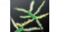
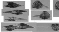
High-resolution imaging allows researchers to examine the intricate morphology and ultrastructure of fungal cells and tissues. SEM provides three-dimensional surface images, revealing details such as cell shape, hyphal branching patterns, spore ornamentation, and surface topography. TEM, on the other hand, offers ultra-thin cross-sections, enabling the visualization of internal cellular components, including organelles, cell walls, and cytoplasmic structures.
2. Fungal Interactions:
Imaging techniques help researchers understand how fungi interact with their environment and other organisms. For example, SEM can capture the interactions between fungal hyphae and host plant tissues during pathogenesis, providing insights into infection mechanisms and disease development. TEM can reveal the ultrastructural details of symbiotic relationships between fungi and beneficial microorganisms, such as mycorrhizal associations in plant roots.
3. Fungal Development and Differentiation:
High-resolution imaging aids in studying fungal development and differentiation. By capturing time-lapse images or serial sections, researchers can observe dynamic processes such as spore germination, hyphal elongation, fruiting body formation, and reproductive structure development. This information is crucial for understanding fungal life cycles and the regulation of developmental processes.
4. Fungal Cell Wall Architecture:
Fungal cell walls are complex structures that play vital roles in growth, protection, and interactions with the environment. High-resolution imaging techniques enable scientists to study the detailed architecture of the cell wall, including its composition, layering, and porosity. This knowledge is essential for understanding fungal biology, pathogenicity, and the development of antifungal agents.
5. Organelle Structure and Function:
TEM allows researchers to investigate the ultrastructure of fungal organelles, including mitochondria, endoplasmic reticulum, Golgi apparatus, vacuoles, and nuclei. By visualizing the structural organization and changes in these organelles during different growth stages or environmental conditions, scientists can gain insights into their functions and contributions to fungal growth and physiology.
6. Nanostructures and Extracellular Matrices:
High-resolution imaging techniques can reveal nanoscale structures and extracellular matrices produced by fungi. These structures play crucial roles in various aspects of fungal biology, such as adhesion, biofilm formation, nutrient acquisition, and communication. Understanding these nanostructures and matrices enhances our comprehension of fungal behavior and ecological interactions.
7. Medical Mycology and Pathogenesis:
In medical mycology, high-resolution imaging is instrumental in studying the morphogenesis of pathogenic fungi, their interactions with host cells, and the mechanisms of infection. This information is vital for developing effective diagnostic tools, understanding virulence factors, and designing antifungal therapies.
In summary, high-resolution imaging techniques offer a window into the intricate world of fungal growth and biology. By providing detailed morphological, structural, and ultrastructural information, these techniques advance our understanding of fungal diversity, physiology, interactions, and applications in various fields, including agriculture, biotechnology, ecology, and medicine.