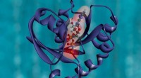
Actin is a thin, flexible protein that forms long filaments. Myosin is a thick, rigid protein that forms motor proteins. Motor proteins are able to move along actin filaments, pulling them towards the center of the cell. This contraction of the actin filaments creates force, which allows the cell to move.
Cells can move in a variety of ways. For example, they can crawl along a surface, they can swim through a liquid, and they can even fly. The type of movement that a cell is able to perform depends on its shape and the arrangement of its actin and myosin filaments.
Cell movement is essential for a variety of cellular processes. For example, cells must be able to move in order to divide, to repair themselves, and to respond to their environment. Cell movement is also important for the development of embryos and for the functioning of the immune system.
Researchers are currently studying how cells move in order to better understand how diseases like cancer and muscular dystrophy develop. By understanding how cells move, scientists may be able to develop new treatments for these diseases.
Here is a more detailed explanation of how cells muster and march out:
1. Cell polarization: The first step in cell movement is cell polarization. This means that the cell establishes a front and a back. The front of the cell is where the cell will move towards, and the back of the cell is where the cell will leave from.
2. Formation of the leading edge: The next step is the formation of the leading edge. The leading edge is a thin, sheet-like protrusion that forms at the front of the cell. The leading edge is made up of actin filaments and myosin motor proteins.
3. Extension of the leading edge: The leading edge then extends forward, pulling the rest of the cell with it. This extension is driven by the polymerization of actin filaments. Actin filaments are added to the leading edge at the front of the cell and then disassembled at the back of the cell.
4. Contraction of the cell body: As the leading edge extends, the cell body contracts. This contraction is driven by the interaction of actin and myosin filaments. Actin filaments are pulled towards the center of the cell by myosin motor proteins.
5. Detachment of the trailing edge: The final step in cell movement is the detachment of the trailing edge. The trailing edge is the rearmost part of the cell. It is detached from the substrate by the action of proteolytic enzymes.
This process of cell movement is repeated over and over again, allowing cells to move around their environment.