Named for the Swiss anatomist, Johann Peyer, who described them in 1677, Peyer's Patches are structures concentrated in the mucosal lining of the ileum, which is the last section of the small intestine. Each patch consists of lymphoid tissue that bulges outward into the intestinal space.
Peyer's Patches interact with antigens found in the intestine to produce antibodies. According to "Physiology of the Gastrointestinal Tract," the intestinal mucosa encounters more antigens than any other body tissue.
Antigens, which are usually proteins or polysaccharides, come from not only harmless food proteins and beneficial bacteria, but from pathogenic bacteria, viruses, and parasites. Antigens contact the Peyer's Patches' epithelial cells, triggering immune responses.
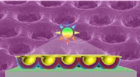
Multifold or M cells are specialized cells in the Peyer's Patches outer epithelial layer. The part of the cell sticking out into the intestine has many tiny folds visible under electron microscopy. These cells take up the antigens and transport them inward to cells that can start antibody formation.
Antigen is transferred to dendritic cells and B and T cells which underly the M cells. B and T cells begin producing antibodies and migrate through the lymphatic system to be released via the thoracic duct into the blood system. Mesenteric lymph nodes and the spleen may also release antibodies when cued from the B and T cells.