The spike protein is a critical component of the virus, and it is the target of many vaccines and treatments. However, the spike protein is also constantly changing, which makes it difficult for scientists to develop effective treatments.
The new visualization technique, called cryo-electron microscopy (cryo-EM), allows scientists to see the spike protein in unprecedented detail. cryo-EM involves cooling the spike protein to very low temperatures and then using an electron microscope to take pictures of it.
The resulting images show the spike protein in its natural state, and they allow scientists to see how the protein moves and changes shape.
This information is critical for understanding how the virus infects cells and how vaccines and treatments can be developed to block the infection.
The researchers published their findings in the journal Nature.
How cryo-EM works
Cryo-EM is a relatively new technique that has been used to study the structure of proteins and other biological molecules. It involves freezing the sample to very low temperatures, which prevents the molecules from moving and allows them to be imaged in their natural state.
The electron microscope used in cryo-EM is much more powerful than a traditional light microscope, and it can produce images with much higher resolution. This allows scientists to see the details of the spike protein in unprecedented detail.
What the researchers found
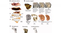
The researchers used cryo-EM to image the spike protein in two different states: the prefusion state and the postfusion state.
The prefusion state is the state in which the spike protein is located on the surface of the virus. In this state, the protein is in a closed conformation, and it is not able to infect cells.
The postfusion state is the state in which the spike protein has fused with the membrane of a host cell. In this state, the protein is in an open conformation, and it is able to inject the virus's genetic material into the cell.
The researchers found that the spike protein undergoes a number of changes in shape as it transitions from the prefusion state to the postfusion state. These changes are critical for the virus to infect cells.
Implications for vaccine and treatment development
The new information about the spike protein's movements could help scientists develop more effective vaccines and treatments for COVID-19.
Vaccines that target the spike protein could be designed to block the protein from changing shape, which would prevent the virus from infecting cells. Treatments that target the spike protein could also be designed to block the protein from fusing with the membrane of host cells.
The researchers hope that their findings will help to accelerate the development of new treatments and vaccines for COVID-19.