A Light microscope, also known as an optical microscope, works based on the principles of magnification and resolution to produce magnified images of tiny structures too small to observe with the naked eye. It involves illuminating a specimen with visible light, allowing magnified viewing through an optical system consisting of lenses in the objective and eyepiece.
Light Microscope Components
A typical light microscope consists of the following main components:
* Objectives: These are sets of lenses located at the bottom of the microscope near the specimen. Multiple objective lenses with different magnifications are usually available on a revolving turret.
* Body (Barrel or Stand): The central structural part of the microscope that supports and connects all major components.
* Stage: The platform where the specimen is placed and prepared for viewing.
* Diaphragm: Located below the stage, it controls the amount of light reaching the specimen.
* Illuminator: A light source, usually a built-in lamp, that provides light for specimen observation.
* Stage Clips: Metal clips used to secure the specimen in place on the stage.
* Eyepiece: The lens or lenses located at the top of the microscope tube, closest to the eye.
* Focusing Knobs: Coarse adjustment and fine adjustment knobs control the vertical movement of the body or the stage to focus on the specimen clearly.
How Light Microscopes Work
The basic operation of a light microscope is as follows:
1. Illumination: Light from the illuminator passes through the diaphragm and the condenser lens, which gathers and directs the light toward the specimen on the stage.
2. Specimen Magnification: The objective lens acts as a primary magnifier, bending (refracting) the light rays coming from the specimen into a real, inverted, and magnified image within the microscope body.
3. Eyepiece Magnification: After passing through the objective lens, the light continues to the eyepiece, where it is further magnified, resulting in a magnified virtual image that appears to originate from the real image formed by the objective.
4. Total Magnification: The total magnification of a microscope is calculated by multiplying the magnification power of the objective lens with that of the eyepiece. For example, using a 40x objective lens and a 10x eyepiece would result in a total magnification of 400x.
Resolution and Contrast
Resolution refers to the ability to distinguish between two adjacent objects in a specimen, while contrast refers to differences in brightness and darkness in the image. These aspects are crucial for obtaining clear and informative microscopic images.
* Resolution: Limited by the wavelength of light used, light microscopes have a resolution range from 0.2 to 2 micrometers (µm). Higher magnification does not always lead to improved resolution.
* Contrast: Several techniques, such as staining, phase contrast, and differential interference contrast, are employed to enhance contrast in light microscopy.
Different Light Microscopy Techniques
Beyond the basic principles described above, various techniques and modifications are employed in light microscopy to study specific types of specimens or enhance imaging capabilities. These include:
* Brightfield Microscopy: The most common technique, it provides bright images against a dark background.
* Darkfield Microscopy: Illuminates the specimen obliquely to produce a dark background and bright objects.
* Phase Contrast Microscopy: Utilizes phase differences in the light to highlight transparent, colorless structures.
* Fluorescence Microscopy: Involves fluorescent dyes or proteins to emit visible light when exposed to specific wavelengths.
Applications of Light Microscopy
Light microscopes are extensively used in research and clinical settings, including:
* Biology: Studying cells, tissues, and microorganisms.
* Microbiology: Examining bacteria, fungi, and protozoa.
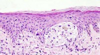
* Pathology: Evaluating tissue samples for diagnosis.
* Forensic Science: Analyzing evidence, including fibers and hair.
* Material Science: Investigating surfaces, particles, and structures of materials.
Light microscopes may not provide the same level of resolution and magnification as electron microscopes but remain indispensable tools across various disciplines due to their ease of use, widespread availability, and ability to observe living specimens under non-destructive visible light.